Most EMT students can name every rhythm on a flashcard and still freeze when the same rhythm appears mid-call. The gap is not knowledge. It is recognition speed.
Flashcards train you to study a frozen strip for twenty seconds; a real call gives you three seconds while someone is talking to you.
This guide covers the twelve rhythms you are tested on, the single giveaway feature of each, and a drill routine that moves recognition from deliberate to automatic.
Why Rhythm Recognition Breaks Down Under Pressure
Recognition fails for a predictable reason: students learn rhythms as static pictures, but rhythms arrive as changes. A patient does not present in ventricular tachycardia, a patient deteriorates into it while you are taking a blood pressure.
If every hour of your study time was spent on still images, the first moving rhythm you see will be on a real patient. This is the gap TrainingMonitor.App was built to close, by letting a rhythm change in front of you the way it does on scene.
The second reason is sequence. Under stress, students reach for the rate first because the number is easy. Rate is the last question, not the first. Regularity and P waves narrow twelve possibilities down to two or three before the rate matters at all.
If reading the full screen is still new to you, work through how to read a vital signs monitor before you start drilling rhythms in isolation.
The 12 Rhythms You Are Tested On
Work through these in order. For each one, learn the giveaway feature the single thing that separates it from everything else.
- Normal Sinus Rhythm: A P wave before every QRS, regular, 60 to 100. This is your reference picture.
- Sinus Bradycardia: The same picture, under 60. Ask whether the patient is symptomatic, not just slow.
- Sinus Tachycardia: The same picture, over 100 and usually under 150. It is a symptom of something else.
- Atrial Fibrillation: Irregularly irregular with no identifiable P waves. If you cannot find a pattern in the spacing, this is your first suspect.
- Atrial Flutter: Sawtooth waves between complexes, often with a fixed conduction ratio.
- Supraventricular Tachycardia: Narrow and fast, roughly 150 to 250, with P waves buried in the preceding T wave.
- First-Degree AV Block: A PR interval longer than 0.20 seconds that stays the same length every beat.
- Second-Degree Type I (Wenckebach): The PR interval stretches a little further each beat until a QRS is dropped.
- Second-Degree Type II: The PR interval never changes, then a QRS disappears with no warning. This one is unstable.
- Third-Degree Block: P waves and QRS complexes each keep their own steady rhythm and ignore each other.
- Ventricular Tachycardia: Wide, fast and regular, with no P waves anywhere.
- Ventricular Fibrillation: Chaotic, with no complex you can measure.
Asystole and pulseless electrical activity sit alongside these, but both are confirmed by the patient rather than the screen. A flat line with a pulse is a lead problem, not asystole.
Three Mistakes That Cost You the Call
The first is reading the rate before the rhythm. Build the habit of asking three questions in fixed order: is it regular, is there a P wave before every QRS, and is the complex narrow or wide. The rate comes fourth.
The second is trusting the number instead of the tracing. A displayed heart rate of 180 can be a patient in SVT or a loose electrode counting motion as beats. The waveform tells you which; the number never does.
The third is practicing only on clean strips. Textbook tracings are printed at the clearest moment. Field tracings have baseline wander, muscle artifact and a patient who moves.
If you can only identify a rhythm when the picture is perfect, you have learned the picture rather than the rhythm.
How to Build a Rhythm Drill Routine
Five focused minutes daily will beat a three-hour session on the weekend. Set a target of naming any rhythm within five seconds, then drill against a clock so the time pressure is part of the practice rather than a surprise on test day.
TrainingMonitor.App is a browser-based vital signs simulator that is enough to run this no mannequin, no hardware and no subscription are required.
Practice transitions, not snapshots. Have a partner change the rhythm without telling you and call out what you see as it happens.
Programs that are still paying for this capability can compare what they are getting against a free alternative to paid ECG rhythm generators before the next renewal comes around.
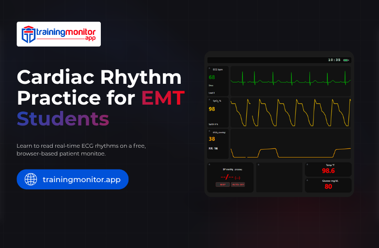
Finally, drill with the whole picture on screen. On TrainingMonitor.App a rhythm runs alongside blood pressure, oxygen saturation and respiratory rate, so you learn what ventricular tachycardia looks like in a patient who is also crashing which is the only way it ever appears on a real call.
Once you can name the twelve reliably, switch seats and become the person generating the rhythm. Running the scenario teaches you how a deterioration actually builds, and on TrainingMonitor.App you can control vital signs in real time while a partner reads them.
Then put the tracing where you will actually see it. Open the display monitor on a second screen, start a rhythm, and practice calling it before the waveform finishes crossing the screen.
Frequently Asked Questions
Q. Which rhythm do EMT students get wrong most often?
Ans. Second-degree type II, because it is mistaken for Wenckebach when the PR interval is not measured across several beats.
Q. How long does rhythm recognition take to learn?
Ans. Most students reach reliable five-second recognition after four to six weeks of short daily drills.
Q. Do I need to memorize exact rates?
Ans. No, learn the boundaries at 60, 100 and 150, and use regularity and P waves to do the rest.
Q. Can I practice without a mannequin or paid equipment?
Ans. Yes, TrainingMonitor.App runs in any browser and generates live, changing rhythms at no cost.